

The ancient anaerobic pathways still at work inside our cells
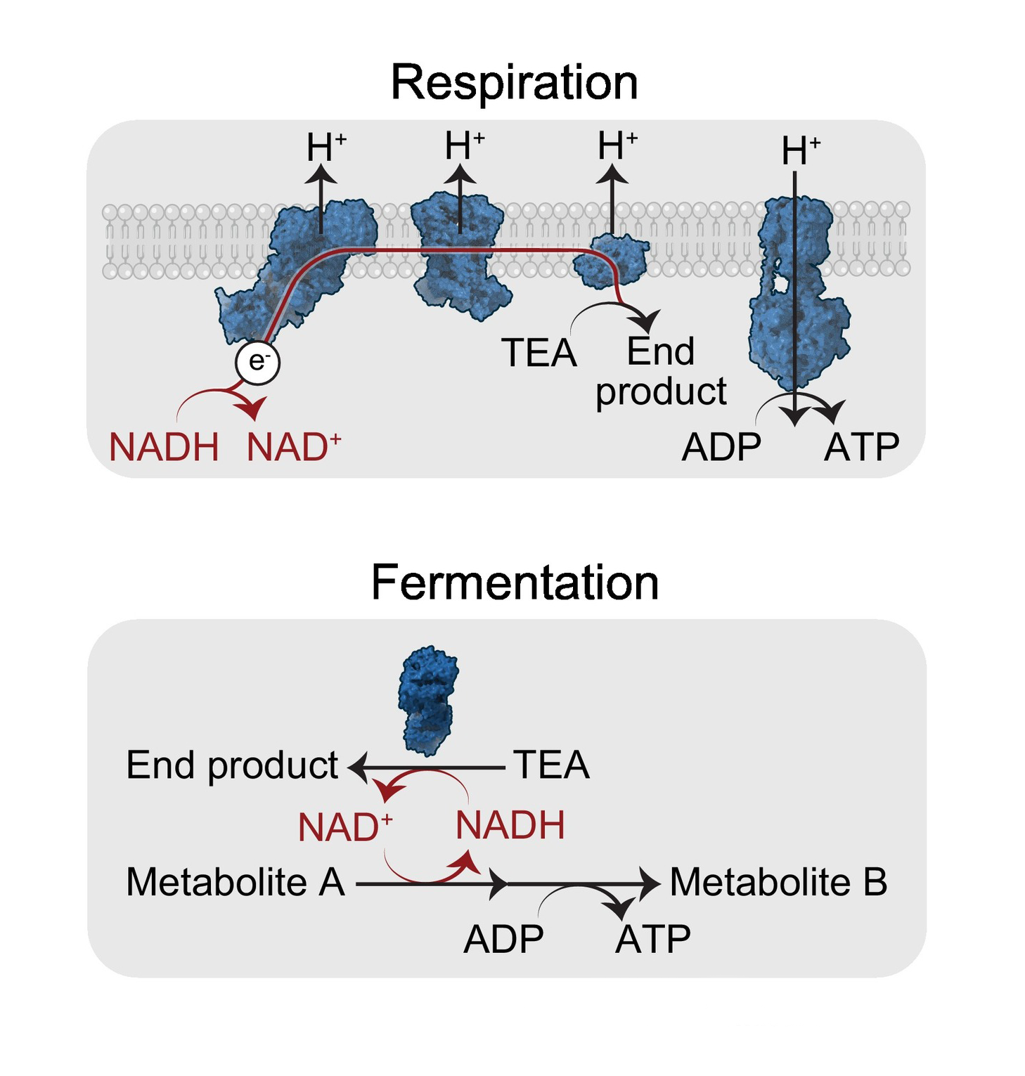
Respiration and fermentation are the two categories of pathways for maintaining electron flow.
Life as we know it depends on the flow of electrons. From single-celled organisms to human neurons, the thermodynamics of this energy flow remain the same: fuel oxidation strips electrons from nutrients, and those electrons move from lower to higher reduction potentials until they come to rest on a terminal electron acceptor (TEA). Without TEAs to clear electrons, NAD+ cannot be regenerated, fuel oxidation stalls, and energy production collapses.
Oxygen is a particularly potent TEA, which is why mammalian energy production relies on it so heavily. But—as we discuss in our review published today in Trends in Biochemical Sciences—the first eukaryotes emerged in an oxygen-poor (i.e. hypoxic) atmosphere. Billions of years ago, before our planet was flooded with oxygen, cells instead relied on anaerobic pathways for energy. Work from our lab is revealing that their descendants, humans included, still retain a repertoire of these anaerobic TEAs.
In hypoxia, the NADH/NAD+ ratio skyrockets inside cells, leading to reductive stress and, eventually, cell death. The canonical fix is the fermentative conversion of pyruvate into lactate, which regenerates NAD+. However, in a preprint published last week, we show that this is not the only cellular response to hypoxia in mammals. We find an ancient strategy for dealing with hypoxia that doesn't involve lactate at all. Instead, it uses metabolites from branched-chain amino acids (BCAAs) as terminal electron acceptors.
In humans, the metabolic end products of BCAA fermentation serve primarily as biomarkers of reductive stress. And, in rodents, we find that BCAA fermentation has evolved for a highly specialized purpose: powering hypermotility in sperm. Across eukaryotes, there is a whole slew of anaerobic strategies for shedding electrons. Perhaps others lie latent in the mammalian metabolic network, available for tuning to specific cellular needs.
Discovering BCAA fermentation in mammals
The starting point for this project was a time- and organ-resolved hypoxia metabolomics dataset that we generated in mice. As we looked through the data for potential electron carriers enriched in hypoxia, we were struck by three metabolites from the branched-chain hydroxyacid (BCHA) family. In every organ at every timepoint, these same three hydroxyacids were enriched in low-oxygen conditions. These weren’t unfamiliar molecules, per se. BCHAs are a well-known product of BCAA fermentation in single-celled organisms, like fermentative bacteria and certain flagellated parasites. What surprised us was finding them enriched in hypoxic mammals. We wanted to know why mice were making them in the first place.
One possibility was that BCHAs enable mice under hypoxia to permanently dispose of excess electrons. This is a documented survival strategy in goldfish, who cope with wintering in oxygen-depleted frozen lakes by increasing ethanol fermentation. As goldfish booze up the lake water by ejecting ethanol from their gills, they also expel the excess of suffocating electrons that ethanol carries along with it. Lacking the gills necessary for this strategy, mammals instead purge unwanted metabolic byproducts through the urinary system. So, we tested the urine of mice for fermentation-related metabolites, and, sure enough, it contained elevated levels of BCHAs. The body seemed to be selectively flushing them out.
We then set out to uncover the source of these BCHAs, expecting to find a rare mammalian enzyme. Instead, GWAS and knockout studies pointed to lactate dehydrogenase A (LDHA), the same enzyme that makes lactate. This created a paradox: if LDHA is designed to make lactate, how and why is it suddenly making BCHAs in the context of hypoxia?
It turns out this was a case of catalytic promiscuity. Rather than being perfectly selective, LDHA can also grab branched-chain ketoacids and reduce them to BCHAs. Under normal conditions, LDHA largely ignores these ketoacids. But, during hypoxia, mitochondria suppress the oxidation of branched-chain ketoacids, and they eventually pile up in the cytosol, forcing LDHA to run the reaction anyway. Hypoxia also drives the accumulation of electrons in the cytosol by impairing respiration, further increasing BCAA fermentation. When we forced electrons to accumulate in the cytosol by blocking the malate-aspartate shuttle, cells started producing BCHAs. We had found the trigger for BCAA fermentation: the cytosolic ratio of NADH/NAD+.
The unique evolution of lactate dehydrogenase in rodent sperm
Because BCAA fermentation increases in hypoxia, we wondered if low-oxygen conditions in humans may also trigger this process. And, indeed, BCHA levels increase in three distinct human states: resistance exercise, in which short bursts of maximal effort require anaerobic ATP production; severe COVID-19, which induces systemic hypoxemia; and alcohol consumption, which leads to NADH accumulation in the liver via alcohol oxidation. Since the pathway exhibits slower flux than lactate production, BCHAs serve primarily as a biomarker of reductive stress in humans.
But we remained puzzled by a separate instance in which BCHAs were most highly elevated: in male vs. female mice. This led us to an understudied isoform of lactate dehydrogenase (LDHC), an enzyme exclusively expressed in the testes. While every rodent LDHC we tested showed high levels of BCAA fermentation, LDHC from non-rodent mammals—including blue whales, lemurs, and humans—did not. It appears that rodent LDHC has uniquely evolved to widen the range of substrates it tolerates, reducing more ketoacids than its counterparts in other mammals.
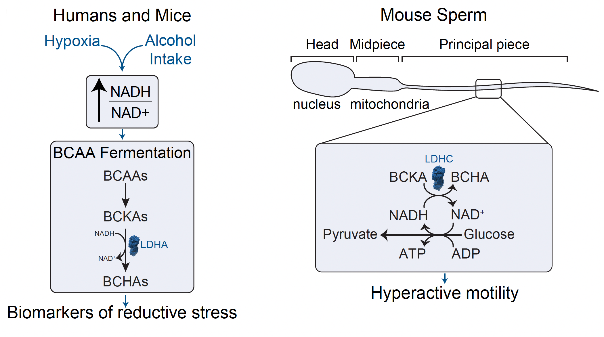
The location of rodent LDHC gives us clues to its utility: these enzymes are wrapped around the motor inside the sperm’s tail, or flagellum, providing energy for sperm to swim rapidly and forcefully towards the egg to fertilize it. It’s not yet clear why rodent sperm evolved towards higher velocities. However, since hyperactivity is a universal requirement across mammals, sperm fermentation could represent a possible therapeutic target in reproductive healthcare.
From a metabolic lens, this discovery highlights that mammalian electron recycling appears to be far more diverse than the pyruvate-to-lactate reaction alone. For over a century, a massive spotlight has focused on lactate as the primary mammalian fermentation pathway. But, by leveraging high-throughput data, we were able to broaden our search space and find another creative way the body responds to the problem of electrons, rooted deep in our biological past. Maybe there are other specialized tissues that harbor their own unique electron sinks. Electrons have to end up somewhere, and, with BCAA fermentation, we’ve identified another place they can go.
Midha, A. D. and Jain, I. H. (2026). The diversity of terminal electron acceptors across eukaryotes. Trends in Biochemical Sciences. https://doi.org/10.1016/j.tibs.2026.07.009
Midha, A. D., Chew, B. T. L., Martí-Mateos, Y., Blume, S. Y., Flanigan, W. R., Desousa, B. R., Haribowo, A. G., Poddar, A., Chadha, S., Queliconi, B. B., Barrios, A. M., Traglia, M., Thomas, R., Suzuki, J., Kuroda, M., Altschuler, S. J., Wu, L. F., Kirichok, Y., Paredes, M. F., Anthony, T. G., Lishko, P. V., & Jain, I. H. (2026). Branched-chain amino acid fermentation as an alternative mammalian electron sink. bioRxiv. https://doi.org/10.64898/2026.08.24.746737
Ayush Midha (X: @AyushDMidha) is an MD/PhD student studying metabolism in the Jain Lab.
Isha Jain (X: @ishahjain) is an Arc Institute Core Investigator, an Associate Investigator at Gladstone Institutes, and an Associate Professor at UCSF.